The suit from 19 states and the District of Columbia contends that major restructuring has cut life-saving programs and put extra burden on states to pay for health crises.
Following Listeria outbreaks and recalls, an ongoing review identified several steps that can be quickly implemented.
Though the funding covers melioidosis and Marburg tests, ASPR said the new capability can be repurposed to develop and produce tests for other biological threats when needed.
As a steady polio rise continues in Pakistan, the country has now reported 45 cases this year.
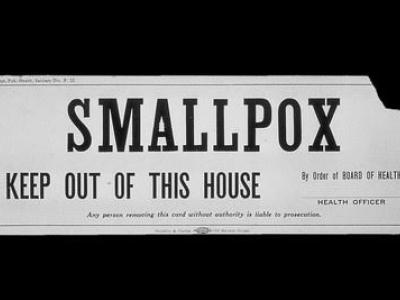
National stockpiles of smallpox vaccines, antivirals, and drugs are "imperfect" and limited, the authors say.
The recommendations include internationally accepted definitions of terminology, greater transparency with the public, and stronger biorisk-management systems and oversight.
COVID vaccine ordering changes on August 3, the US destroys its last chemical weapons, and Mandy Cohen takes the CDC reins.
The low incidence of pathogens with bioterrorism potential would hamper efforts to isolate them from natural sources for intentional release, the authors say.
Opening of the NBAF in Kansas marks the culmination of an effort since 2006 to replace the 68-year-old Plum Island Animal Disease Facility in New York.
In the lab breach, virus was detected in a sewage sample and was genetically similar to vaccine stocks used at the facility, then a lab worker tested positive.