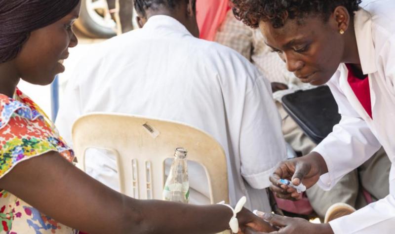
Global health groups say evolving developments, including a novel reassortant in Asia and increasing detections in mammals, require real-time monitoring.
The monoclonal antibody, L9LS, was previously shown to offer 80% protection to adults in a phase 1 trial.
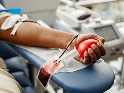
Adults with long COVID were about twice as likely to experience depression, anxiety, sleep difficulties, cognitive problems, and disabling fatigue.
Infectious disease information from CIDRAP.
Discussion & analysis on the latest infectious disease developments by Dr. Osterholm and Chris Dall.
CIDRAP staff are frequently featured on other news sites. Please see our opinion pieces, commentaries, and other features.
Data on novel vaccine candidates in clinical or late preclinical development.
CIDRAP and Osterholm Update podcast merchandise is available for purchase. Order t-shirts, mugs, socks, buttons, stickers, and more!
The Influenza Vaccines R&D Roadmap Initative
The Influenza Vaccines Roadmap provides a much-needed framework for organizing the efforts of existing influenza researchers while identifying a wide range of opportunities that will encourage new investigators to join the work.
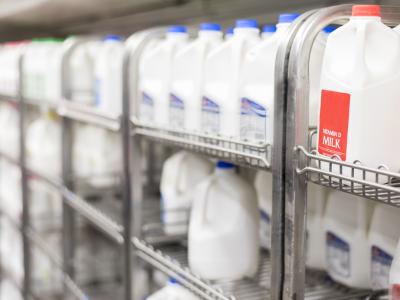
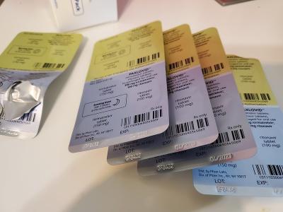
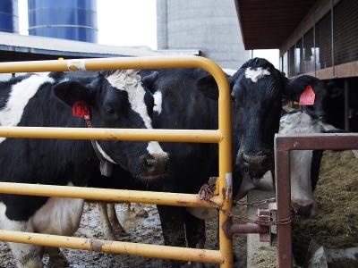
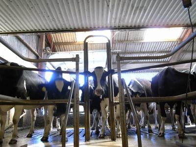
“I wouldn’t have any problem drinking milk tonight from an [H5N1] influenza standpoint at all. My grandchildren could drink the milk tonight.”
Study on masks vs N95 respirators for health workers spurs concerns
The findings suggest medical masks may offer similar protection as respirators against COVID-19, but experts say hold on.