Children and teens with long COVID have significant lung abnormalities detected with an advanced form of magnetic resonance imaging (MRI), called free-breathing phase-resolved functional lung (PREFUL) MRI. The findings were published yesterday in Radiology.
Though chest scans are used to diagnose and monitor lung function of adults with long COVID, they are less commonly used on children with persistent symptoms after COVID-19, which is also called post-COVID condition (PCC). Lung perfusion, or how blood flows in and out of the lungs, can thus be hard to detect in pediatric patients.
Imaging without contrasts or radiation
"Because of radiation exposure and the necessity for intravenous contrast agent, CT [computed tomography] is not used as a standard diagnostic tool in children suspected of having PCC," the authors wrote. "Instead, pulmonary function tests and echocardiography, along with a comprehensive review of medical history, are performed to rule out pulmonary and cardiac differential diagnoses. However, these examinations frequently show normal lung and cardiac function in symptomatic patients, complicating the diagnostic process."
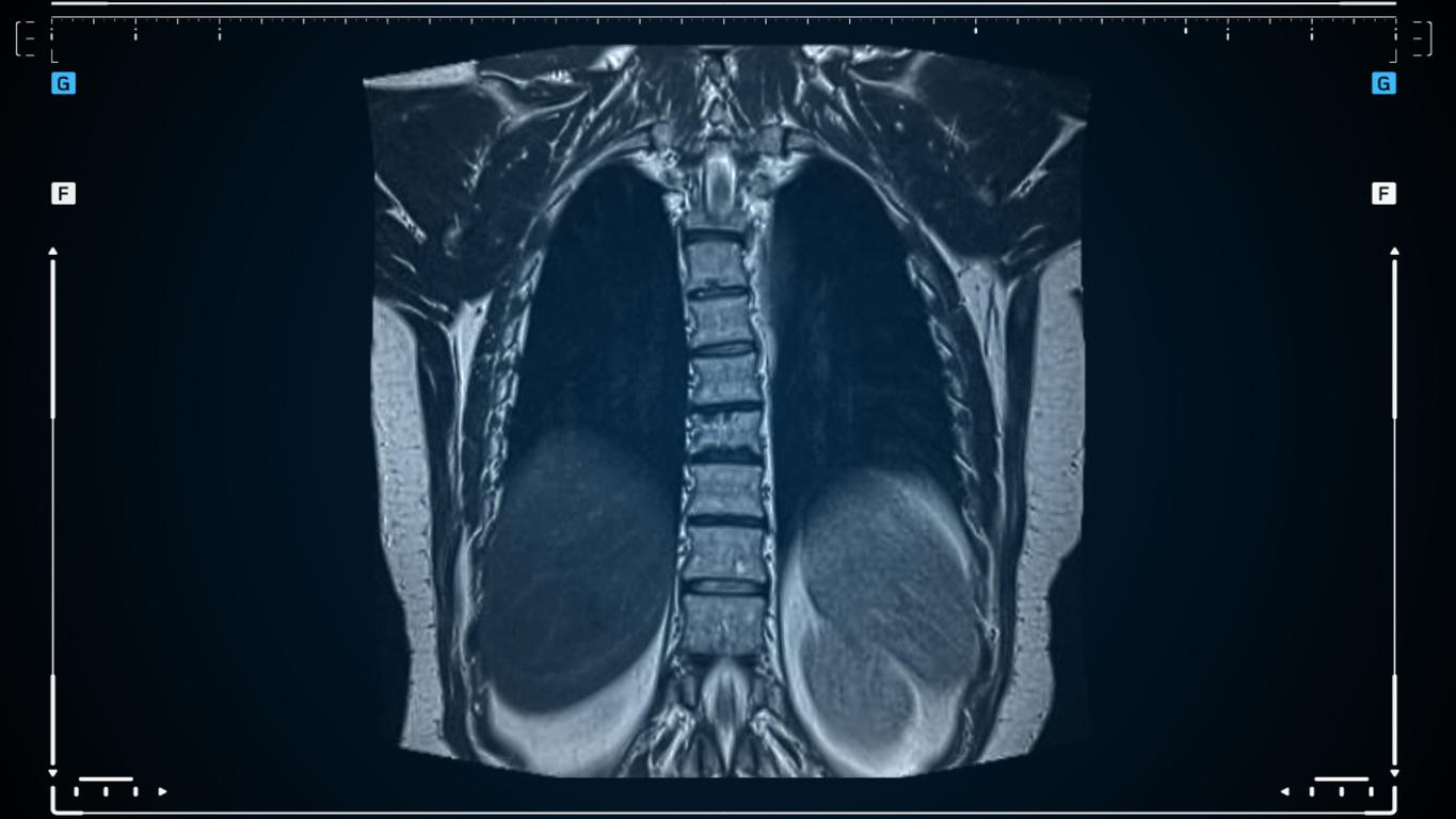
PREFUL MRI, however, is a contrast agent–free and radiation-free imaging modality that may be more suitable for children.
In this study, 54 participants (27 with long COVID and 27 without) underwent PREFUL from April 2022 to April 2023. The imaging assessed regional ventilation (oxygen flow), flow-volume loop correlation metric (FVL-CM), quantified perfusion (blood flow), ventilation and perfusion defect percentages, and ventilation-perfusion ratios.
The median age of participants was 15 years.
Lung injuries linked to chronic fatigue
The PREFUL results showed that kids with long COVID had lung injuries that correlated to specific long-COVID symptoms and overall loss of blood flow in the lungs. In participants with long COVID, greater lung perfusion correlated with increased chronic fatigue severity. In addition, higher ventilation-perfusion mismatch correlated with increased heart rate.
"Our research provides the first comprehensive evidence of measurable regional lung perfusion abnormalities in pediatric post-COVID-19 condition using radiation-free, contrast-free lung imaging," said Gesa H. Pöhler, MD, a senior physician at the Hannover Medical School in Germany and first author of the study, in a news release from the Radiological Society of North America, which publishes the journal.
The authors said PREFUL could also be used on long-COVID patients to determine how or if they are improving.
Quantitative lung MRI establishes a potential imaging biomarker profiling and helps to enable disease severity follow-up for this complex condition in the future.
"Quantitative lung MRI establishes a potential imaging biomarker profiling and helps to enable disease severity follow-up for this complex condition in the future," Pöhler said.
The authors concluded, "Further investigations should prioritize multicenter longitudinal studies with larger cohorts to validate these findings and evaluate the progression of lung abnormalities at various stages after COVID-19 infection."