Patients with long COVID may exhibit persistent inflammation in the heart and lungs for up to 1 year following acute COVID-19 infection, even when standard medical tests return normal results, according to a new study in the Journal of Nuclear Medicine.
The study authors, from the Icahn School of Medicine at Mount Sinai, suggest the inflammation may increase the risk for future cardiac and pulmonary conditions.
This study brings us closer to understanding how SARS-CoV-2 affects the heart and lungs over time.
"This study brings us closer to understanding how SARS-CoV-2 affects the heart and lungs over time. We believe long COVID results in an inflammatory response that may predispose patients to premature coronary artery disease, pulmonary hypertension, and valvular damage such as stenosis or regurgitation," said first author Maria G. Trivieri, MD, PhD, in a press release from Mount Sinai.
57% had evidence of inflammation
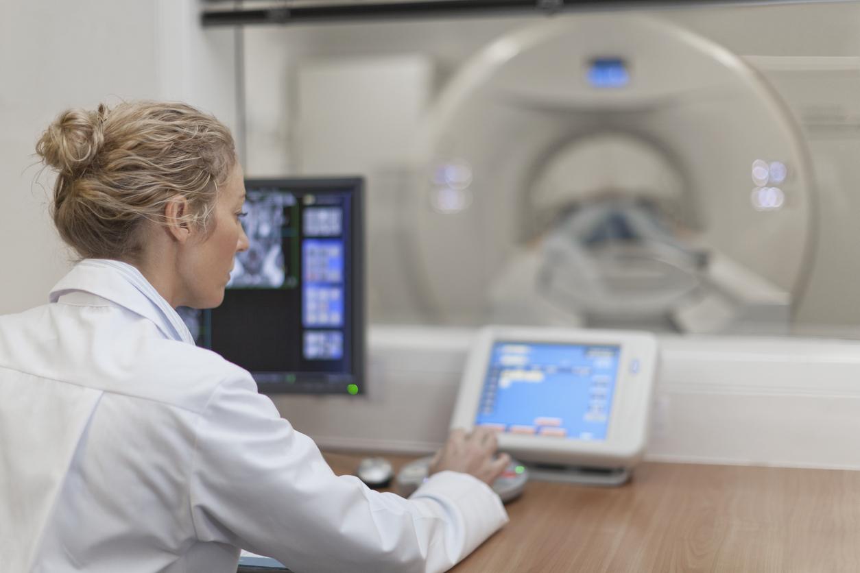
To conduct the study, the researchers performed PET/MRI (positron emission tomography and magnetic resonance imaging) scans on 99 patients who reported persistent cardiopulmonary symptoms 9 to 12 months after confirmed COVID-19 infection in 2020 and 2021. Eighty percent of patients had shortness of breath (80%), and 27% of participants were hospitalized.
Images were compared to a control group of nine participants with a history of acute severe COVID-19 infections but no lasting symptoms. The imaging was done on average 300 days after their initial infections.
Overall, 57% of participants had evidence on PET/MRI of inflammation affecting the heart muscle, pericardium (the thin sac that surrounds the heart), heart valves, particularly the mitral valve, and the aortic and pulmonary blood vessels on imaging. The most common abnormality, seen in 28 participants, was vascular inflammation involving the aorta or pulmonary arteries. Twenty-two patients had myocardial (heart muscle) scarring and thickening of tissue.
None of the control group had signs of inflammation.
"This study highlights the unique power of hybrid PET/MRI imaging to uncover hidden disease processes in long COVID patients," said Zahi Fayad, PhD, senior author of the study.
"These findings should change how we approach care and surveillance—not only recognizing SARS-CoV-2 as a potential long-term cardiovascular risk factor, but also integrating molecular imaging into post-COVID evaluation protocols," Fayad added. "We now have objective evidence that can guide earlier detection and potentially prevent future cardiopulmonary events."