Jan 27, 2012
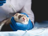
In ferrets, H5N1 can reach nervous system via olfactory mucosa
A Dutch research team has found that an H5N1 avian influenza virus can spread to the central nervous system (CNS) of ferrets via the olfactory mucosa, according to a report in the Journal of Virology. Intranasal inoculation of ferrets with a wild-type H5N1 strain led to extensive growth of the virus in the olfactory muscosa, from where it spread to the olfactory bulb of the brain and the rest of the CNS, the report says. The finding shows that "the olfactory epithelium forms an island of susceptible cells in a sea of relatively resistant respiratory epithelium." The virus also spread to the heart, liver, pancreas, and colon, suggesting it traveled through the blood as well. The team also determined that systemic spread of the virus depended on the presence of the multibasic cleavage site in the virus's hemagglutinin, as ferrets inoculated with an H5N1 strain lacking this site had infections only in the respiratory tract. The researchers say that most flu viruses in mammals are confined to the respiratory tract, but H5N1often spreads systematically. They write that further studies should aim to determine if flu viruses other than highly pathogenic H5N1 can invade the CNS via the olfactory route.
Jan 25 J Virol abstract
Australia reports first low-path avian flu outbreak
Australia's agriculture ministry today reported the country's first known low-pathogenicity avian influenza outbreak in poultry, which turned up during routine surveillance on a commercial duck farm near Melbourne, according to a report today from the World Organization for Animal Health (OIE). Sequencing tests confirmed that the virus is a low-pathogenicity H5 subtype. Polymerase chain reaction tests have ruled out N1 involvement, but more tests are being done to further characterize the virus. The birds on the farm appeared healthy. Culling of 24,500 birds to curb the spread of the virus included ducks at a second duck-breeding farm that had business links to the first farm, the OIE report said. An investigation hasn't found the source of the virus.
Jan 27 OIE report
Study tracks viral co-evolution in real time
In experiments with the phage lambda virus and Escherichia coli bacteria, researchers from Michigan State University demonstrated how adaptation by natural selection plays a role in virus evolution, according to a EurekAlert press release. The findings appear today in the latest issue of Science. The group identified a novel tactic the virus used to infect its host, involving mutations on the virus's host-recognition protein. They observed that after the bacteria reduced expression of the original receptor, LamB, virus mutations allowed infection through a new receptor, OmpF. Justin Meyer, a study coauthor, said in the press release that his group was surprised to see the lambda virus gain the new function so quickly. In a Science podcast, he said the group replicated the experiment 96 times and saw the virus evolve to develop the novel pathway 24 times. He said that on average it took the virus 12 days to evolve the new function. "What we found is that the reason why it was so fast, and the reason why it happened so many times is that natural selection was really driving the evolution and driving the fixation of the mutations responsible for this new function," he added. In an accompanying perspective piece, John N. Thompson of the department of ecology and evolutionary biology at the University of California, Santa Cruz, wrote that the experiments show that co-evolution can play a key role in shaping innovation. He commented that the experiments occurred in ecologically simple environments and that futher research is needed to explore if the changes are more or less likely in more variable environments.
Jan 26 EurekAlert press release
Jan 27 Science abstract
Jan 27 Science Perspective summary
Jan 27 Science podcast
E coli acquired in Turkey similar to 2011 European outbreak strain
A French woman who returned home from a tour of Turkey last September with hemolytic uremic syndrome (HUS) was infected with a strain of Shiga toxin–producing Escherichia coli (STEC) O104:H4 similar to the one that caused a widespread outbreak in France and Germany earlier in 2011, according to a report in Eurosurveillance. The earlier outbreak was attributed to contaminated fenugreek sprouts. The French traveling group comprised 22 people; eight experienced diarrhea, including two hospitalized with HUS on their return. No common food source could be traced, but no one in the group reported eating sprouts before or during the trip. The timing of the cases suggested the illnesses were acquired in Turkey. Both HUS cases showed positive serology for STEC O104:H4. One, which showed the same resistance profile as the strain in the European outbreak, was compared by pulsed-field gel electrophoresis with cases from the earlier outbreak. It was similar but not identical, showing differences in two bands for the gene Xbal and in three bands for Notl. The authors conclude from these and several cases that occurred from 2004 through 2009 that STEC O104 circulates in Turkey and certain other countries, such as Egypt and Tunisia, and that the strain should be considered in travelers who return from these areas with HUS.
Jan 26 Eurosurveillance article